
When back pain or neck pain begins to interfere with daily life, pinpointing the cause is the first step toward relief. For many patients, that process starts with imaging — a crucial tool that helps physicians see what’s happening beneath the surface.
At Texas Spine & Neurosurgery Center, board-certified neurosurgeon Dr. Rajesh Bindal combines more than two decades of experience with advanced imaging technology to accurately diagnose spine disorders. His expertise in minimally invasive spine surgery, artificial disc replacement, and other precision-based treatments allows him to develop personalized care plans that address the root cause of each patient’s pain.
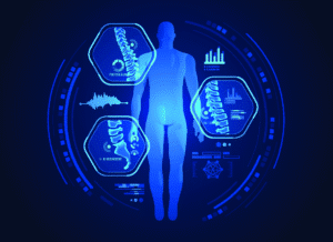
The spine is a complex structure of bones, discs, nerves, and soft tissue. Even a minor abnormality can cause significant discomfort or nerve irritation. Imaging tests such as MRI, CT scans, and X-rays help identify structural changes that may be compressing or inflaming nerves, causing pain to radiate through the arms, legs, or back.
Accurate imaging allows Dr. Bindal to:
- Confirm or rule out conditions such as herniated discs, spinal stenosis, or degenerative disc disease.
- Determine whether surgery or non-surgical treatment is the best approach.
- Map out precise surgical plans for faster recovery and minimal disruption to surrounding tissue.
Common Imaging Techniques for Spine Conditions
- Magnetic Resonance Imaging (MRI): Often considered the gold standard for spine evaluation, MRI provides detailed images of the discs, spinal cord, and nerves without radiation. It’s especially helpful for diagnosing herniated discs, nerve compression, and soft tissue changes.
- Computed Tomography (CT) Scans: CT scans use X-rays to create cross-sectional views of the spine. They offer excellent bone detail and can reveal fractures, spinal alignment issues, or bone spurs contributing to nerve pressure.
- X-Rays: X-rays are a fast, cost-effective tool for identifying bone-related issues like arthritis, scoliosis, or vertebral instability. Though less detailed than MRI or CT, they often serve as the first imaging step in a diagnostic plan.
- Myelography: In select cases, a contrast dye is used during imaging to better visualize nerve roots and spinal cord structures, helping diagnose complex nerve compression cases.
Beyond Diagnosis: How Imaging Guides Treatment
Imaging isn’t just about finding the problem — it’s about planning the right solution. For patients considering spine surgery, imaging helps Dr. Bindal visualize the exact area that requires treatment and tailor a surgical approach that minimizes recovery time and scarring.
For patients seeking conservative care, imaging may reveal opportunities to avoid surgery altogether by identifying conditions that respond well to physical therapy, medications, or targeted injections.
Advanced, Patient-Centered Spine Care
At Texas Spine & Neurosurgery Center in Sugar Land, Dr. Rajesh Bindal integrates cutting-edge imaging with evidence-based treatment to ensure accuracy at every stage of care. The philosophy is simple: when diagnosis is precise, treatment is more effective, and recovery is smoother. Whether you’re struggling with back pain, numbness, or radiating discomfort, Dr. Bindal provides the expertise and advanced tools needed to help you move confidently again. Call 281-313-0031 to schedule a consultation.